| 目录: 28355 |
| 产品名称: CD138(DM56) Rabbit Monoclonal Antibody |
| 基因符号: SDC1; Syndecan-1; CD138; SYND1; SDC |
| 描述: CD138 antibody(DM56) Rabbit Monoclonal Antibody |
| 背景: Syndecan-1 (SYND1 or SDC1) is also known as CD antigen CD138; is a transmembrane (type I) heparan sulfate proteoglycan and is a member of the syndecan proteoglycan family. The syndecans mediate cell binding; cell signaling; and cytoskeletal organization and syndecan receptors are required for internalization of the HIV-1 tat protein. The syndecan-1 : SDC1 protein functions as an integral membrane protein and participates in cell proliferation; cell migration and cell-matrix interactions via its receptor for extracellular matrix proteins. It is a useful marker for plasma cells; but only if the cells tested are already known to be derived from blood. |
| 经过测试的应用: ELISA; Flow Cyt |
| 推荐稀释度: ELISA 1:5000-10000; Flow Cyt 1:100 |
| 种属反应性: Rabbit |
| 亚型: Rabbit IgG |
| 纯化: Purified from cell culture supernatant by affinity chromatography |
| 种属反应性: Human CD138 |
| 成分: Lyophilized from sterile PBS, pH 7.4. 5 % – 8% trehalose is added as protectants before lyophilization. |
| 储存和运输: Store at -20°C to -80°C for 12 months in lyophilized form. After reconstitution, if not intended for use within a month, aliquot and store at -80°C (Avoid repeated freezing and thawing). |
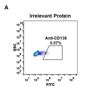 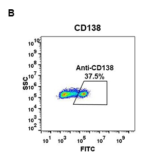
Figure 1. Expi 293 cell line transfected with irrelevant protein (A) and human CD138 (B) were surface stained with Rabbit CD138 monoclonal antibody 1µg/ml ( clone: DM56) followed by Alexa 488-conjugated rabbit IgG secondary antibody. | 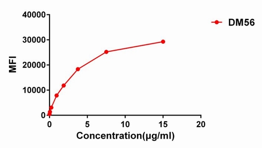
Figure 2. Flow cytometry data of serially titrated Rabbit CD138 monoclonal antibody ( clone: DM56) on H929 cells. The Y-axis represents the mean fluorescence intensity (MFI) while the X-axis represents the concentration of IgG used. | 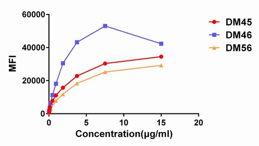
Figure 3. Affinity ranking of different Rabbit CD138 mAb clones by titration of different concentration onto H929 cells. The Y-axis represents the mean fluorescence intensity (MFI) while the X-axis represents the concentration of IgG used. | |
|